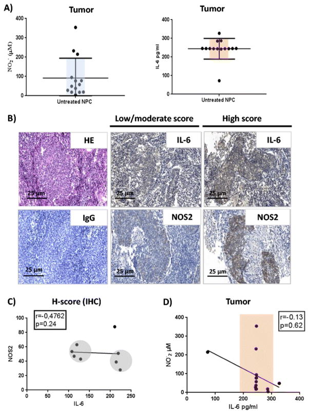
Fig 2. Analysis of IL-6 and NOS2 signals in NPC patient’s tumors.
A) Analysis of nitrites and IL-6 levels in biopsy supernatants of untreated patients cultured biopsies (n=14). The horizontal lines show the Mean ± SD of the groups. B) IL-6 expression NOS2 expression in untreated NPC patients’ biopsies. The tumor nests and the tumor immune infiltrating cells express IL-6 with more intensity in the stroma (Magnification x20). NOS2 is expressed by tumor and by the immune cells infiltrating the tumor microenvironment (Magnification x20). HE stainings and isotype control antibody IgG labelling were used as negative controls. C) Correlation between IL-6 and NOS2 staining in NPC patients (H-score). A negative correlation was observed between NOS2 and IL-6 among patients (n=8; r=−0.47). D) Correlation analysis between NO2- and IL-6 concentrations in NPC tumor explants supernatants. No correlation was found between nitrites and IL-6 among NPC patients (n=12) (Spearman’s test coefficient r=0.13).

