
Figure 4.
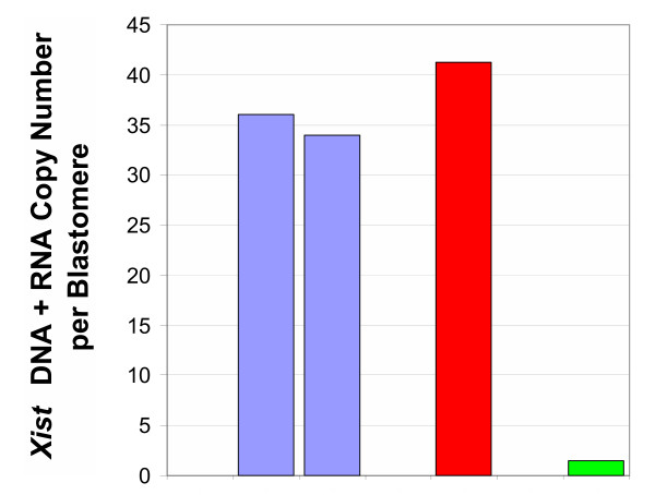
Quantification of Xist RNA levels in isolated blastomeres and whole embryos at the 4-cell stage. Comparison of Xist genomic DNA + cDNA (RNA) levels measured in single blastomeres isolated from a 4-cell female embryo and in whole 4-cell embryos of either sex. Whole embryo data are presented on a per cell basis. Light purple bars, two individual blastomeres harvested from the same female embryo. Red bar, whole female embryo (per blastomere average). Two Xist template copies in each female cell are accounted for by the presence of Xist genomic DNA, while the remaining copies signal the presence of Xist RNA. Green bar, whole male embryo (per blastomere average): as expected, the number of Xist copies in this sample corresponds to the presence of one copy of the Xist gene per cell and the absence of Xist transcripts. Sexing was confirmed in all samples by the presence or absence of the Sry gene.