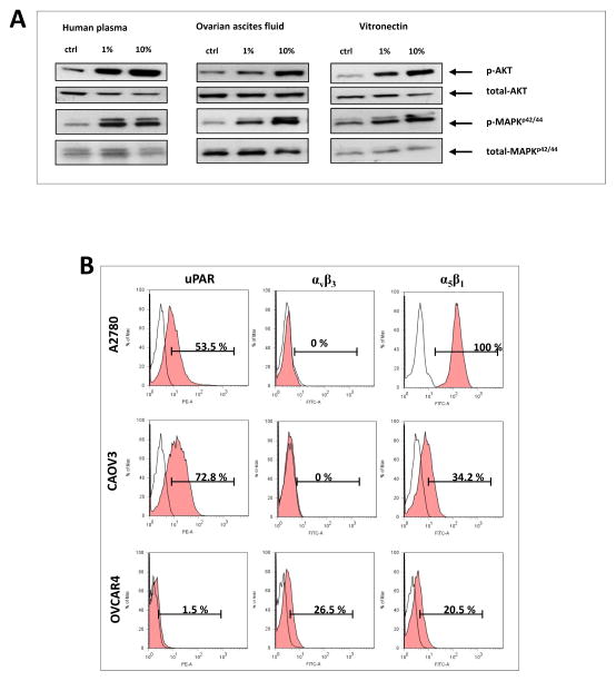
Figure 3. Ovarian cancer cell lines express functional vitronectin receptor.
Panel A. Phosphorylation of p42/44 MAPK and AKT after stimulation of A2780 cells with human plasma, ovarian ascites and vitronectin. Experiment was performed twice with similar results. Panel B. Flow cytometry analysis of the expression of urokinase receptor (uPAR) and the two integrin receptors αvβ3 and α5β1 in different ovarian cancer cell lines. Flow cytometry analysis was performed at least twice for each of the cell lines.

