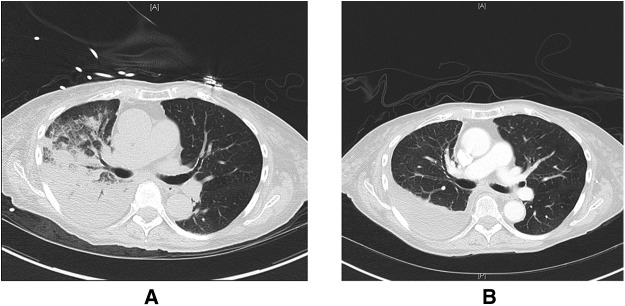
Figure 3.
Representative images from computed tomography scans of the chest before and after therapy for reexpansion pulmonary edema. (A) Ground-glass opacities and airspace disease in the right lung after thoracentesis consistent with reexpansion pulmonary edema. (B) Resolution of these abnormalities and a residual right-sided pleural effusion after diuresis and supportive care.

