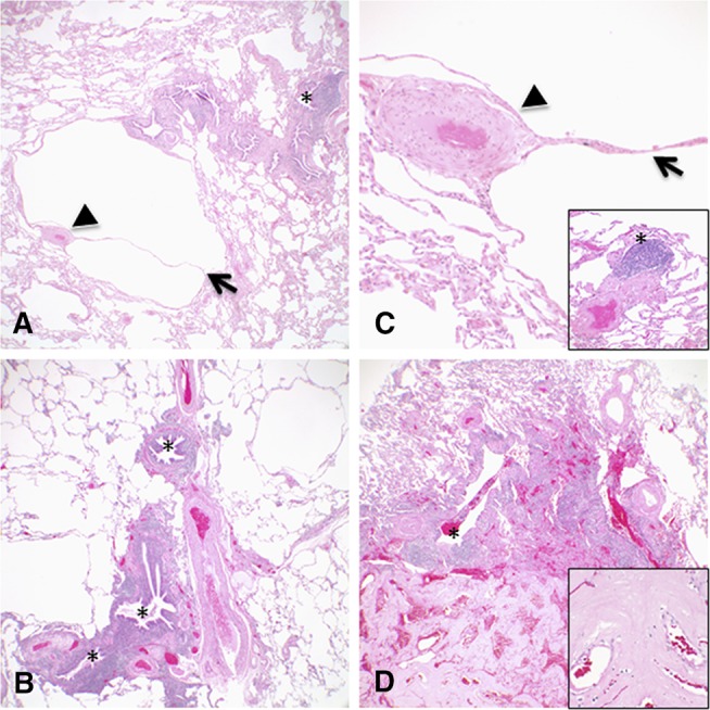
Figure 3.

(A–D) Representative histologic images corresponding to the radiographic features showing multiple varying sized parenchymal cysts, some associated with eccentric vessels (arrowheads) and internal septations (arrows). Follicular bronchiolitis was present in the lung biopsies from all three patients characterized by lymphoid hyperplasia with reactive germinal centers cuffing bronchioles (*). One lung biopsy also had focal subpleural nodular amyloidosis diagnosed by accumulations of amorphous eosinophilic material that stained deep pink to red with Congo red stain and showed apple green birefringence with polarized light (D, bottom of image and inset). Original magnifications: 4× (A, B, and D); 20× (C, including inset); 40× (D, inset).
