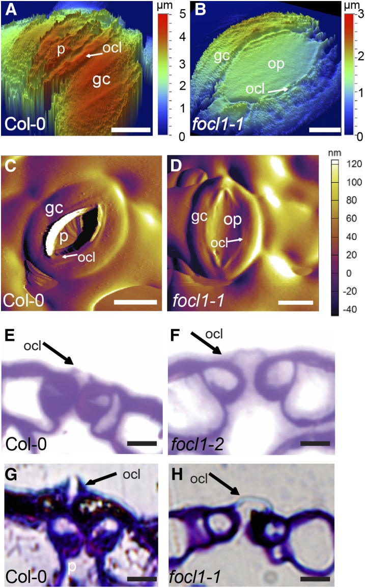
Figure 3.
focl1 mutants have fused outer cuticular ledges. A and B, Abaxial surfaces of wild-type Col-0 and focl1 stomates imaged by VSI. Depth is indicated in nanometers. C and D, AFM deflection images of stomates. E and F, Transverse sections of stem epidermis stained with Toluidine blue. Position of outer cuticular ledges (ocl) indicated by arrows. G and H, Adaxial leaf epidermis. Bars = 5 μm in A, B, E, and F and 10 μm in C, D, G, and H. p, Stomatal pore; ocl, outer cuticular ledge; gc, guard cell; op, occluded pore.

