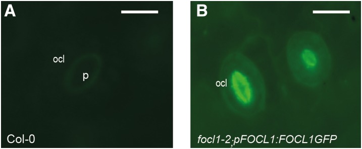
Figure 5.
FOCL1-GFP localizes to the cuticular ledge. Seedlings of T2 lines of focl1-2 expressing pFOCL1:FOCL1-GFP were analyzed by epifluorescence microscopy. Wild-type Col-0 samples showed weak autofluorescence (A) compared to complemented focl1-2 plants (B) where FOCL1-GFP signal is largely restricted to the OCL in developing (right) and mature guard cell (left). Bar = 15 µm.

