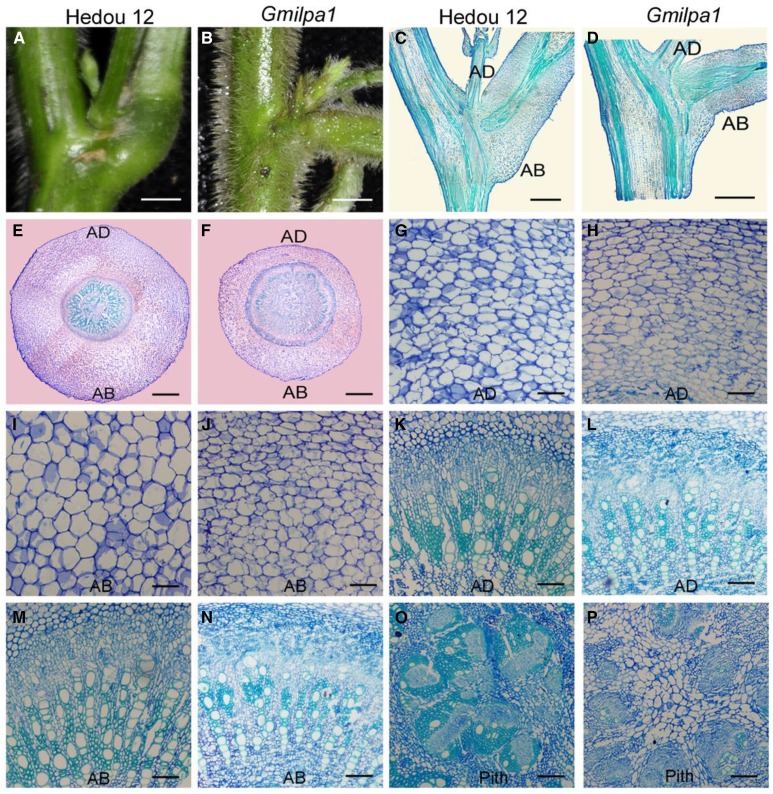
Figure 2.
Structural comparison of pulvini of 6-week-old Hedou 12 and the Gmilpa1 mutant. A and B, Pulvini of Hedou 12 (A) and the Gmilpa1 mutant (B), respectively. Scale bars, 1 cm. C and D, Longitudinal section of pulvini in Hedou 12 (C) and the Gmilpa1 mutant (D), respectively. Scale bars, 1 cm. E and F, Transection of pulvini in Hedou 12 (E) and the Gmilpa1 mutant (F), respectively. Scale bars, 400 μm. G to P, Partial magnification of transection of pulvini in Hedou 12 (G, K, and O) and the Gmilpa1 mutant (H, L, and P). Scale bars, 50 μm. G and H, The motor cells of Hedou 12 (G) and the Gmilpa1 mutant (H) on the adaxial side. I and J, The motor cells of Hedou 12 (I) and the Gmilpa1 mutant (J) on the abaxial side. K and L, The vascular cylinders of Hedou 12 (K) and the Gmilpa1 mutant (L) on the adaxial side. M and N, The vascular cylinders of Hedou 12 (M) and the Gmilpa1 mutant (N) on the abaxial side. O and P, The piths of Hedou 12 (O) and the Gmilpa1 mutant (P).

