Figure 4.
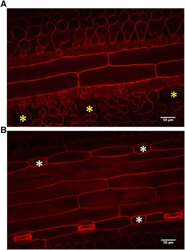
Cellular structure of HvEPF1OE stomatal complexes. A, Representative propidium iodide-stained confocal image of a Z-plane below the HvEPF1OE-1 abaxial epidermal surface. Yellow asterisks mark the location of the substomatal cavity under mature guard cells. B, Higher Z-plane image of the same field of view as A to reveal position of stomata. White asterisks mark the location of arrested stomatal precursors and the lack of underlying substomatal cavities in A.

