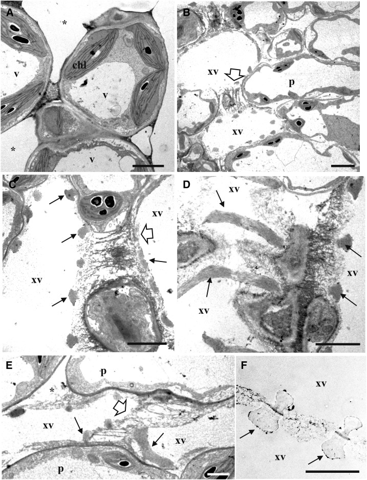
Figure 2.
Observations of the epithem of cauliflower hydathodes by transmission electron microscopy. Ultrathin paradermal sections were stained with PATAg to visualize polysaccharides. A, The epithem, below the epidermal layer, is composed of vacuolated (v) parenchyma cells (p) with chloroplasts (chl) containing heavily stained starch granules. Note the presence of large intercellular spaces (*). B to E, Visualization of xylem vessels (xv) within the epithem. Each xylem vessel exhibit annular thickenings (arrows). A loose fibrillar matrix is observed between adjacent xylem vessels (white arrow) and between vessels and adjacent epithem cells (*). F, A comparable fibrillar matrix is also observed between xylem vessels outside the hydathode tissue. Scale bars = 3 µm.

