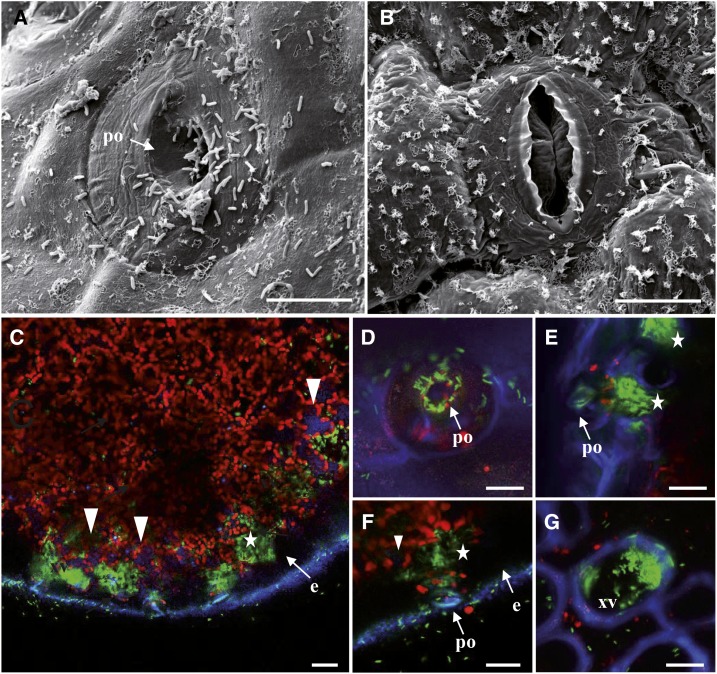
Figure 5.
Cauliflower infected by Xcc strain 8004::GUS-GFP. A and B, Typical scanning electron micrographs of a pore at the hydathode’s surface (A) and a stomata at the leaf surface (B) 3 dpi by transient dipping in the bacterial suspension. Scale bars = 10 µm. A, Note the presence of a large number of bacteria rods inside the hydathode pore (po) and at the surface of the epidermal layer. B, Bacteria are not observed near stomata of the leaf blade. Note the numerous wax ornamentations on the epidermis. C to G, Confocal images of cauliflower infected by GFP expressing bacteria at 3 dpi (C–F) and 6 dpi (G). Images are maximal projections computed from 15 to 25 confocal planes acquired in the z dimension (increment of 0.5 µm). Scale bars = 20 µm. C, Paradermal optical section (parallel to the epidermis) of an infected hydathode at 3 dpi. GFP-labeled bacteria are mainly located in large pockets (white stars) beneath the epidermis (e). Note the absence of bacteria within the epidermal cells (e) and the faint blue autofluorescence inside some parenchyma cells (white arrowheads). D, Visualization of GFP-labeled Xcc at the hydathode pore level (comparable to scanning electron micrograph in A). E to F, Detailed localization of GFP-labeled bacteria in large pockets (white stars) beneath the pore (po) of the hydathode. G, Detection of GFP-labeled bacteria in a xylem vessel (xv) in a transversal section of the mid rib.

