Figure 3.
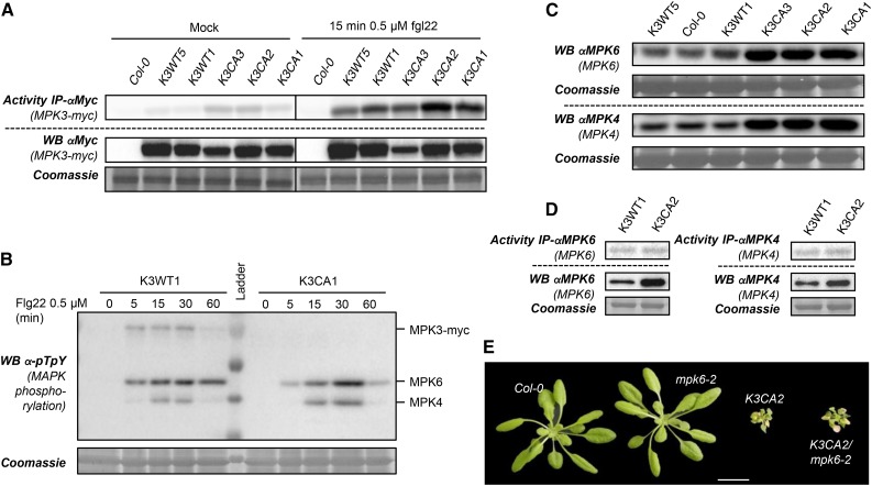
Characterization of MAPK protein levels and PAMP-triggered immunity in K3WT and K3CA lines. A, Kinase activity of MPK3-myc after immunoprecipitation (IP) using anti-c-myc antibody from in vitro plantlets of the indicated genotypes treated with flg22 for 15 min. Western blots (WB) show protein expression levels. B, Kinase phosphorylation in the MAPK activation loop monitored by western blot using anti-pTpY antibody from K3WT1 and K3CA1 transgenic lines treated with 1 µm flg22 for the indicated times. Note that the anti-pTpY antibody does not detect the phosphorylated MPK3 activation loop when it is mutated (D193G/E197A). C, Western blot analyses of MPK4/6 levels using MAPK-specific antibodies in rosettes of pot-grown plants of the indicated backgrounds. D, Kinase activity of MPK4 and MPK6 after immunoprecipitation using specific antibody from rosettes of the indicated genotypes. Western blots show protein expression levels. E, Representative photographs of 1-month-old K3CA2/mpk6-2 plants compared with parental lines and Col-0. Plants were cultivated at 22°C on soil in a culture chamber. Bar = 2 cm.

