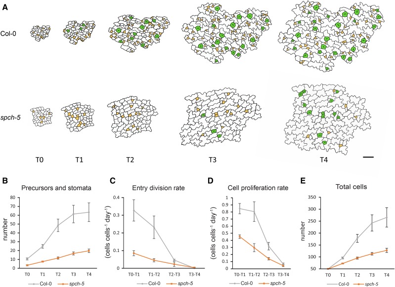
Figure 2.
In vivo tracking of epidermal cell divisions and stomatal differentiation in leaf primordia. A, Abaxial epidermis of third-leaf primordia in Col-0 and spch-5 plants followed in vivo with serial resin imprints. Epidermal replicas were inspected at 24-h intervals for 4 d (T0–T4). Drawings are reproductions from representative micrographs of the Col-0 and spch-5 serial imprints at the times indicated. Stomatal precursors are marked in yellow and stomata in green. B to E, Graphs of the number of stomata and stomata precursor cells (B), entry division rate (C), cell proliferation rate (D), and number of total cells (E) from leaves (n = 5) per genotype and with 50 cells at the initial field (T0) of 50 cells. Error bars represent se. Bars = 100 µm in A.

