Figure 3.
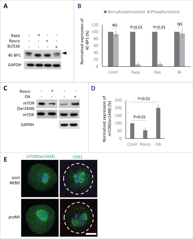
Protein kinases phosphorylating 4E-BP1 in the oocytes. (A) Detection of 4E-BP1 by immunoblotting in the oocytes treated with specific inhibitors Rapa (100 nM), Rosco (10 µM), or BI2536 (100 nM) post-NEBD. Arrowhead marks the presence of upper band (phosphorylation shift) of 4E-BP1 in the oocytes treated for 2 h post-NEBD, GAPDH was used as a loading control, a typical experiment from at least 3 replicates is shown. (B) Quantification of non-phosphorylated and phosphorylated form of 4E-BP1 in the post NEBD oocytes. Data are presented as mean ± SD, Student's t-test, NS = non-significant. (C) CDK1 effect on mTOR phosphorylation in the oocytes treated by Rosco (10 µM) or OA (1 µM). Immunoblot was probed with mTOR(Ser2448) and control (mTOR and GAPDH) antibodies. Twenty oocytes were used per sample. (D) Presence of mTOR(Ser2448) normalized to the mTOR in the Rosco or OA treated oocytes. Data are presented as mean ± SD, Student's t-test. (E) Localization of mTOR(Ser2448) and CDK1 in the post NEBD and pro-MI stage oocytes, n ≥ 30, phospho-specific antibody (green) and DNA (blue). Scale bar = 20 µm.

