. 2017 Apr 27;5(2):S0068. doi: 10.5702/massspectrometry.S0068

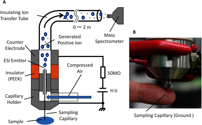
Fig. 1. (A) Schematic of the self-aspirated direct sampling ESI source. The sampling capillary is held at ground potential for the safety of the operator as well as for in-vivo and in-situ mass spectrometry. The electrospray is initiated by applying a high negative potential to the counter electrode for detection in the positive ion mode. The ions and charged droplets are transported to the inlet of the mass spectrometer by the carrier gas from an air compressor. (B) Photograph of the fabricated ion source.

Fig. 1. (A) Schematic of the self-aspirated direct sampling ESI source. The sampling capillary is held at ground potential for the safety of the operator as well as for in-vivo and in-situ mass spectrometry. The electrospray is initiated by applying a high negative potential to the counter electrode for detection in the positive ion mode. The ions and charged droplets are transported to the inlet of the mass spectrometer by the carrier gas from an air compressor. (B) Photograph of the fabricated ion source.