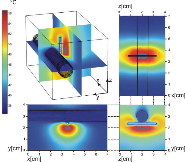
Figure 5.
The heat distribution and the damage in the volume. Left: the heat colour index in °C. The damage appears when the value of the damage function Ω (eq. 8) reaches the threshold of 0.6. Here the image shows the results after 200 s. The damage zone is shown in grey. Fig. 5 is available also as a video stream demonstrates the temperature rise inside the tissue. The video stream shows where, how, and when this damage appears (see additional file 1).

