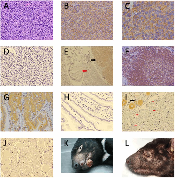
Fig 1. DFT1 staining and skin manifestation.
(A) Haematoxylin and Eosin stained DFT1 x40, (B) ERBB3 Immunohistochemical expression in DFT1 strain 3 x40, (C) ERBB3 immunohistochemical expression in DFT1 strain 3 x100, (D) DFT1 negative control, (E) Tasmanian devil skin and subcutis section with peripheral nerve (red arrow) and DFT1 (black arrow) x10, (F) Tasmanian devil lymph node ERBB3 expression lymphoid follicle x20, (G) Tasmanian devil bowel ERBB3 positive control x40, (H) ERBB3 IHC negative control bowel, (I) trigeminal nerve shows ERBB3 positive nerve body (black arrow) and occasional adaxonal ERBB3 positivity (red arrows) x40, (J) ERBB3 IHC negative control trigeminal nerve, (K) Tasmanian Devil gross appearance of DFT1. Photo credit: DPIPWE archive, (L) Tasmanian devil gross appearance cutaneous lymphoma. Photo credit DPIPWE archive.

