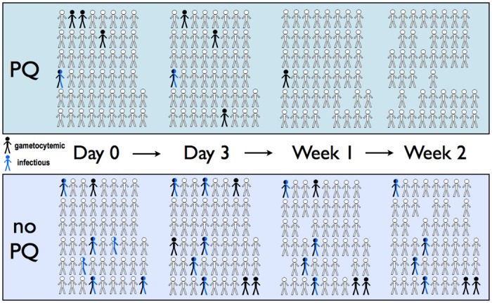
Fig 1. Schematic of gametocyte and mosquito infectivity status through treatment in 101 randomized participants.

Participants in the primaquine (PQ) and non-primaquine arms are depicted in the same ordered configuration from Day 0 pre-treatment through Week 2 post-treatment. Subjects with patent gametocytes detected by microscopy are colored black, while subjects who infected at least one mosquito on membrane feeding are colored blue. Persons that were both gametocytemic and infectious are colored half black-half blue. Persons who missed follow-up are shown as missing.
