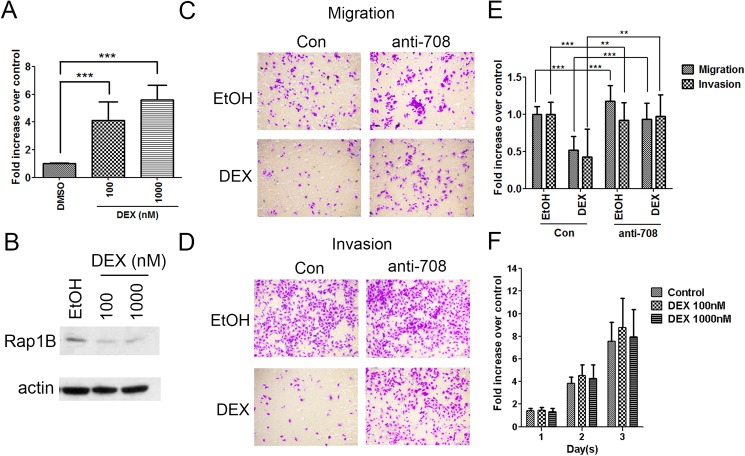
Fig 3. GC signaling reduces the migration and invasion abilities of ID-8 cells through miR-708 induction.
(A) miR-708 expression in ID-8 cells after 72 h of treatment with an increasing amount of DEX (100–1000nM). Data are normalized mean ± SD from three independent experiments. The level of DMSO control group was normalized to one. ANOVA followed by Dunnett's post-hoc test was used for the statistical test (***P < 0.001). (B) Western blot analysis showing Rap1B expression in ID-8 cells upon treatment with 100–1000nM DEX for 48 h. β-actin was used as the loading control. (C–E) ID-8 cells transfected with anti-miR-708 or a control were treated with 1000nM DEX overnight and subsequently subjected to migration (6 h) and invasion (16 h) assays. Representative images of the migration (C) and invasion (D) assays are shown. (E) The cells in three random microscopic fields (200×) were enumerated for each group. Data are normalized mean ± SD combined from three independent experiments. The percentage of migration/invasion obtained for control group was normalized to one. ANOVA followed by Dunnett's post-hoc test was used for the statistical test (**P < 0.01; ***P < 0.001). (F) Cell proliferation assay was performed in ID-8 cells by MTS reagent. 1x10³ cells were cultured and assayed for proliferation in 96 wells. In the next day, ID-8 cells were treated with 100–1000nM DEX for 3 days. Data are normalized mean ± SD combined from three independent experiments. Data presented as fold change from untreated (day 0) expression level.

