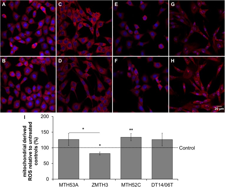
Fig 6. Influence of 10 mM DCA on mitochondrial activity in mammary cell lines after 48 hours.
(A-H) Fluorescence of mitochondrial derived ROS (red) and counterstained cell nuclei (DAPI, blue). (I) In comparison to untreated control significant improvement of mitochondrial activity was observed in mammary carcinoma cell line MTH52C. A visible but insignificant increase was observable in the mammary carcinoma cell line DT14/06T and non-cancerous mammary gland derived cell line MTH53A. The adenoma cell line ZMTH3 showed significantly decreased mitochondrial activity in comparison to untreated control and non-cancerous cell line MTH53A. No significant difference was evaluated between MTH53A and the other cell lines. Data are shown as mean ± SD, n≥3 and are presented as relative fluorescence (mitochondrial activity) in comparison to untreated control (%). Control was set to 100%. Statistical analysis was performed with two-tailed t-test, *p<0.05, **p<0.01, ***p<0.001. (A) MTH53A control; (B) MTH53A+DCA; (C) ZMTH3 control; (D) ZMTH3+DCA; (E) MTH52C control; (F) MTH52C+DCA; (G) DT14/06T control; (H) DT14/06T+DCA; (I) relative mitochondrial activity.

