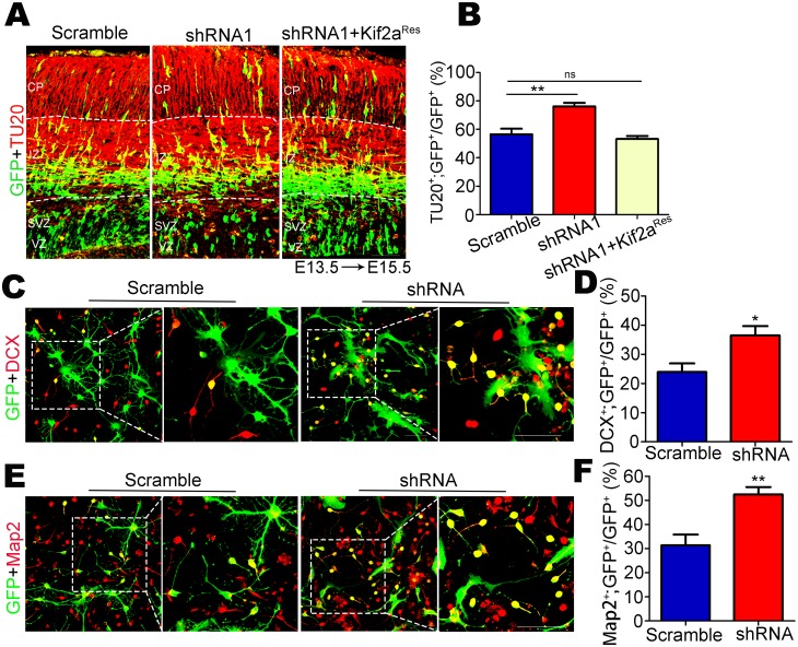
Fig 5. Kif2a is involved in the differentiation of NSCs/NPCs.
(A) Mouse embryos were electroporated with indicated plasmids (scramble shRNA, Kif2a shRNA1 or Kif2a shRNA1+Kif2aRes) at E13.5, and analyzed at E15.5. GFP (green) represents cells expressing the indicated plasmids; TU20 (red) represents immature neurons. Scale bars = 50 μm. (B) Quantitative analysis of the percentage of GFP+;TU20+ cells among the total GFP+ cells showed that more GFP-labeled TU20+ neurons were located in IZ and CP layers after knockdown of Kif2a. n = 800–1000 cells from three different brains. **P < 0.01, ns = no significant difference; one-way ANOVA followed by Bonferroni post-hoc test. Data are presented as the mean ± SEM. (C, E) Cultured NSCs/NPCs transfected with lentivirus expressing scramble shRNA or Kif2a shRNA were incubated in differentiation medium for 3 days. Co-immunostaining for GFP (green) with the neuronal marker DCX (red) in (C) or Map2 (red) in (E). Scale bars = 50 μm. (D, F) Quantification of the percentage of GFP+;DCX+ or GFP+;Map2+ cells among total GFP+ cells showed that knocking down Kif2a increased the number of newborn neurons. n = 600–800 cells from three different experiments. *P < 0.05; **P < 0.01; Student’s t-test. Data are presented as the mean ± SEM.

