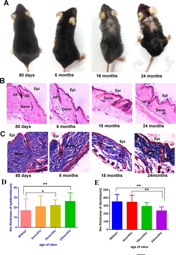
Fig 1.
Representative images of mice of different ages, 80 days mice (n = 6), 6 months mice (n = 6), 18 months mice (n = 6), 24 months mice (n = 3) (A). HE staining of the skin from different ages mice (B,C), For HE and Masson staining, representative images from 8–16 tissue sections in 2–6 mice are shown the thickness of full skin and epidermis of different age mice(D,E).Data are expressed as the mean ± s.e.m. *** P<0.005, unpaired t-test, two-tailed. Scale bars, 50 μm.

