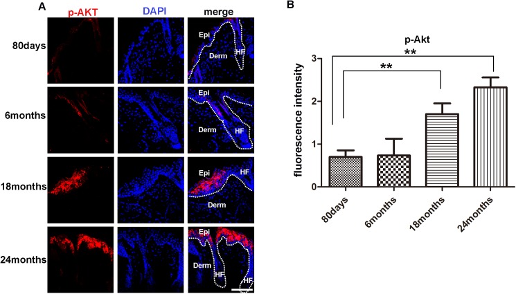
Fig 3. Levels of p-Akt in the skin of mice in different ages.
(A)(IF?)Immuno-fluorescence analysis barely detected the presence of p-Akt positive cells in the skin of 80 days and 6 months old mice, but detected evident p-Akt positive cells in the epidermis and hair follicle of 18 and 24 months old mice. For all IF analyses, representative images from 8–16 tissue sections in 3–6 mice are shown. (B) Fluorescence intensity of p-Akt of 80days, 6months, 18 months, 24months old mice were measured by image J. Scale bars, 20 μm for microscopic images. Data are expressed as the mean ± s.e.m. *** P<0.005, unpaired t-test, two-tailed.

