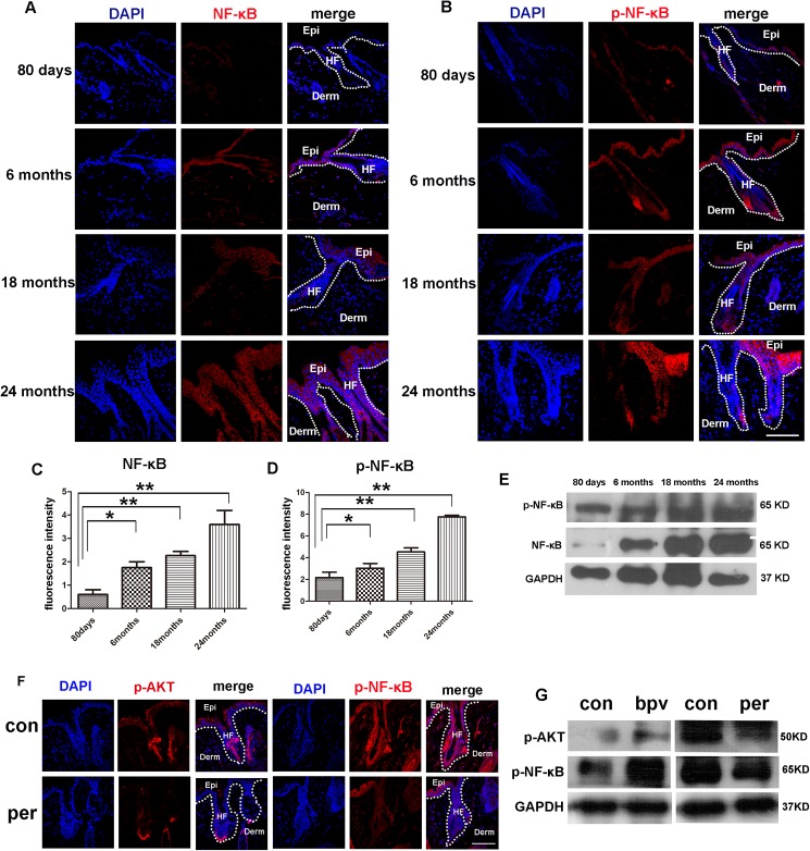
Fig 4. Levels of NF-κB, p-NF-κB in the skin of mice in different ages.
(A, B) Immuno-fluorescence analysis showed increasing levels of NF-κB and p-NF-κB in the skin of mice with aging. Scale bars, 20 μm. (C,D) Fluorescence intensity of NF-κB, p-NF-κB of 80days,6months,18months, 24months old mice were measured by imageJ. (E) Western blotting analysis showed the levels of NF-κB and p-NF-κB in the skin tissue of mice aged 80 days, 6 months, 18 months and 24 months. (F) Immuno-fluorescence analysis showed increasing levels of p-AKT and p-NF-κB in the skin of 12 months mice treated with perifosine and the littermate without perifosine treated (N = 3). Scale bars, 20 μm. (G) Western blotting analysis showed the levels of p-AKT and p-NF-κB in the skin of 12 months mice treated with perifosine and 80 days mice which treated with the bpv(phen), the littermate no-treated mice as control group (N = 3).

