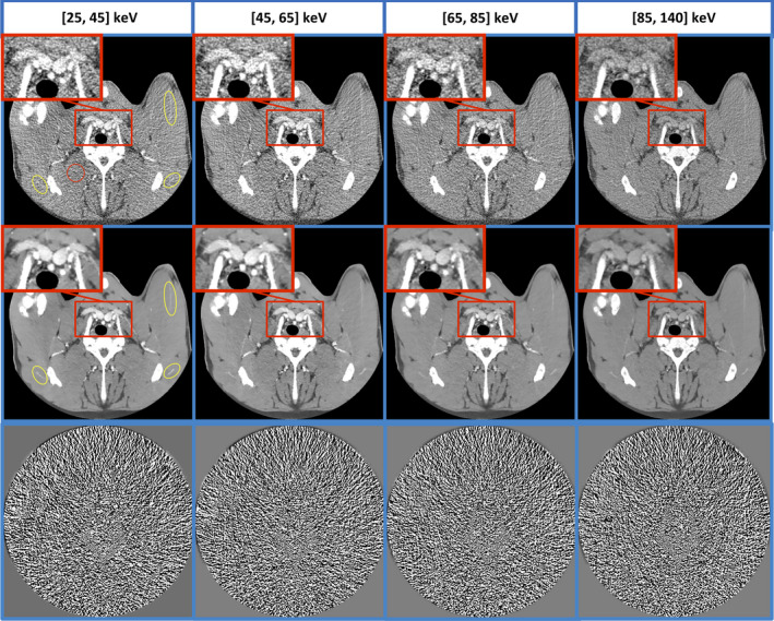
Figure 12.

Bin‐based images after MENLM filtering (middle row, W/L = 400/40 HU) improved the detection of subtle enhanced vessels white ROIs in low‐energy bin‐based image) and low‐contrast structures (Close‐ups in high‐energy bin‐based image). In original FBP images (top row, W/L = 400/40 HU.), the mean and standard deviation of CT number inside the red ROI are 62.2 ± 55.6, 62.8 ± 57.8, 67.2 ± 48.8, and 57.9 ± 32.5, respectively. After MENLM filtering, the values are 62.7 ± 9.1, 62.6 ± 10.8, 65.7 ± 9.5, and 57.7 ± 7.2, respectively. No obvious image structures or signal bias were observed in the difference images (W/L = 15/0). [Color figure can be viewed at wileyonlinelibrary.com]
