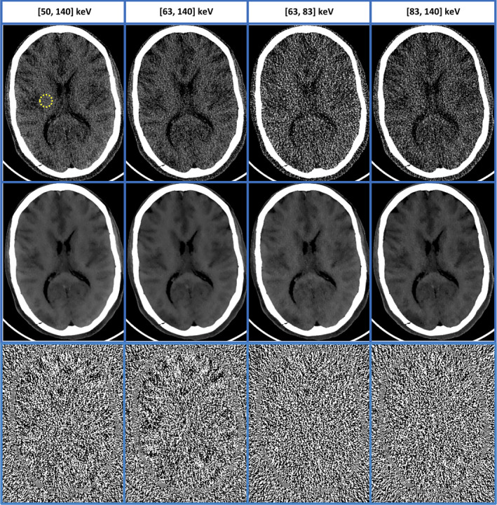
Figure 13.

MENLM filtering of two noisy threshold‐based and two bin‐based images (middle row, W/L = 150/20 HU) from a cadaver head scan demonstrated improved low‐contrast resolution. The similarity/weight determined from threshold‐based images provided robustness to noise reduction. (In original FBP images (top row, W/L = 150/20 HU), the mean and standard deviation of the CT number in the dotted ROI were −10.7 ± 15.1 HU, −12.5 ± 18.2 HU, −9.8 ± 40.6 HU, and −13.3 ± 31.1 HU, respectively. With MENLM, the values were −11.3 ± 2.4 HU, −13.1 ± 2.3 HU, −10.0 ± 3.7 HU, and −14.3 ± 3.8 HU, respectively.) No obvious image structures or signal bias were observed in the difference images (W/L = 15/0). [Color figure can be viewed at wileyonlinelibrary.com]
