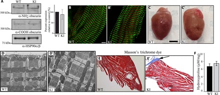
Fig. 1. Examination of the hearts of 1-year-old homozygous female knock-in mice under sedentary conditions.

(A) Immunoblotting using antibodies to the NH2 and COOH termini of obscurins and protein lysates prepared from left ventricles of wild-type (WT) and knock-in (KI) hearts indicated similar expression levels of wild-type and mutant obscurins. Please note that obscurin A is the major giant isoform expressed in the heart, which is shown in our blots, whereas obscurin B is present in low amounts and is only detectable after longer exposure. The expression levels of obscurin A were quantified and found to be unaltered; n = 3 animals per group, two to three replica blots per heart; t test, P = 0.629; error bar represents SEM; heat shock protein 90 (HSP90)α/β served as loading and normalization control. (B and B′) Immunofluorescence combined with confocal optics and antibodies to the COOH terminus demonstrated that mutant obscurins containing the R4344Q substitution are incorporated normally into sarcomeric M-bands in knock-in left ventricles; obscurins are shown in red, and α-actinin, which is a Z-disk marker, is shown in green; scale bar, 5 μm. (C and C′) Gross morphology of wild-type and knock-in hearts showed that they have comparable sizes; scale bar, 3 mm. (D and D′) Transmission electron micrographs of wild-type and knock-in left ventricles revealed no major structural alterations; scale bar, 500 nm. (E and E′) Masson’s trichrome staining of wild-type and knock-in left ventricle sections illustrating the presence of peripheral fibrosis (arrow) in the latter; scale bar, 100 μm. (F) A trend of increased hydroxyproline content was found in the apices of knock-in left ventricles compared with wild-type tissue, which, however, did not reach statistical significance (t test, P = 0.202; n = 4 animals per group, three replica assays per heart).
