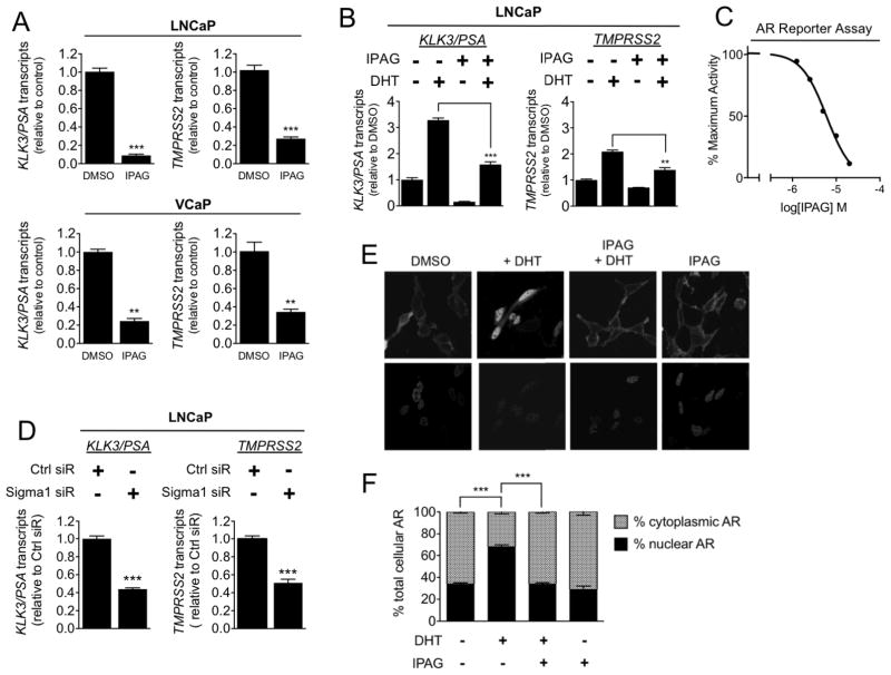
Figure 2. Small molecule Sigma1 inhibitor suppresses AR transcriptional activity and DHT-induced AR nuclear translocation.
(A) Detection of KLK3/PSA and TMPRSS2 mRNA transcripts by qRT-PCR from LNCaP and VCaP cells treated for 16 hours with 20 μM IPAG under steady-state, normal culture conditions. (B) Detection of KLK3/PSA and TMPRSS2 mRNA transcript levels by qRT-PCR from LNCaP cells grown in CSS medium for 72 hours then subjected to co-treatment of 1 nM DHT with DMSO or 10 μM IPAG for 3 hours. (C) AR transcriptional activity luciferase reporter assay. Dose-related inhibition of androgen agonist, 6α-Fl Testosterone (6α FlT) stimulated AR transcriptional activity by IPAG (2.5, 5, 10, 20, 40 μM), 6 μM IC50. (D) Detection of KLK3/PSA and TMPRSS2 mRNA transcript levels by qRT-PCR from LNCaP cells wherein Sigma1 was knocked down by siRNA. Immunoblot confirmation of Sigma1 knockdown in LNCaP shown in Fig. 4A. Data are presented as mean ± S.E.M from at least three independent experiments. (E) Confocal micrographs of LNCaP(GFP-AR) cells grown in charcoal-stripped medium for 3 days followed by 3 hour treatment with 1 nM DHT alone or in the presence of 10 μM IPAG. (F) Quantification of relative nuclear and cytoplasmic GFP-AR levels. For each condition, at least 5 cells were counted per field in at least 5 fields from 3 independently performed experiments, and data are presented as mean ± S.E.M. **P < 0.01; ***P < 0.001.

