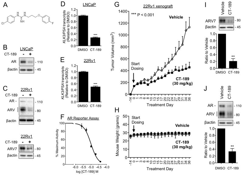
Figure 7. In vivo efficacy: Small molecule Sigma1 inhibitor suppresses growth of ARV driven CRPC tumor xenografted mice.
(A) Chemical structure of 1-(4-chlorophenyl)-3-(3-(4-fluorophenoxy)propyl)guanidine (CT-189). (B) Immunoblots of AR in LNCaP cells treated for 16 hours with CT-189 (20 μM). (C) Immunoblots of AR, ARV, and ARV7 from 22Rv1 cells, grown in CSS medium, treated for 16 hours with CT-189 (20 μM). (D–E) Detection of KLK3/PSA mRNA transcript levels by qRT-PCR from LNCaP cells (D) and 22Rv1 cells grown in CSS medium for 5 days (E), then subjected to 16 hour treatment with 20 μM CT-189. (F) Luciferase reporter assay showing dose-responsive inhibition of AR transcriptional activity by CT-189 (1.25, 2.5, 5, 10, 20, 40 μM). (G) In vivo tumor growth inhibition by CT-189 in castrated mice xenografted with 22Rv1 cells. Treatment with CT-189 was started 14 days post-implantation of 22Rv1 cells, when tumors reached ~150 mm3. Either drug vehicle or CT-189 (30 mg/kg) was administered by oral gavage every other day (q.a.d., quaque altera die). N ≥ 8 for each treatment arm. One-way analysis of variance (ANOVA) was used to determine statistical significance, ***P < 0.001. (H) Mouse weight (grams) during treatment course. No significant weight loss compared to vehicle was observed in CT-189 (30 mg/kg) treated mice. (I–J) Tumors were measured and harvested within 48 hours after final drug administration, at treatment day 36. Tumor proteins were extracted and immunoblotted to evaluate changes in ARV7 (I) and AR (J) protein levels. Note that in panel J only AR was quantified. Bars represent mean ± S.E.M. and are generated from at least three tumors for each treatment condition analyzed in triplicate. **P < 0.01; ***P < 0.001.

