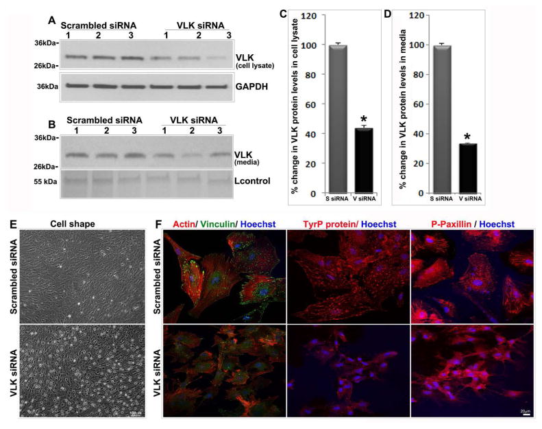
Figure 4.
VLK deficiency in human TM cells induces changes in cell shape and decreases in actin stress fibers and focal adhesions. TM cells (lanes 1–3 represent 3 individual samples) treated with siRNA specific to VLK for 72 h showed a significant decrease in VLK protein levels in both cell lysates (A & C) and media (B & D) compared to the control cells treated with scrambled siRNA, based on immunoblotting and densitometric analyses. Values are mean ± SEM of 12 independent analyses. *P<0.05. Lower panels in A & B show the loading controls. E. TM cells treated with VLK siRNA for 72 h reveal changes in cell shape by phase contrast microscopy compared to the control cells treated with scrambled siRNA. F. TM cells treated with VLK siRNA exhibit a decrease in actin stress fibers (Rhodamine-phallodine staining), focal adhesions (vinculin staining) and in phospho-tyrosine and phospho-paxillin immunofluorescence compared to control cells treated with scrambled siRNA. Bars in E & F show image magnification.

