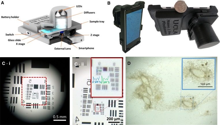
Figure 1.
(A) Schematic demonstrating the design of our mobile phone–based microscope. (B) Photos of the mobile phone microscope. (C) Quantification of spatial resolution using U.S. Air Force (USAF) test resolution chart. (c-i), an image of USAF chart taken using the mobile microscope; (c-ii), zoomed in region of USAF chart shown in c-i. (D) An image of Schistosoma haematobium eggs on the membrane captured using our mobile phone–based microscope.

