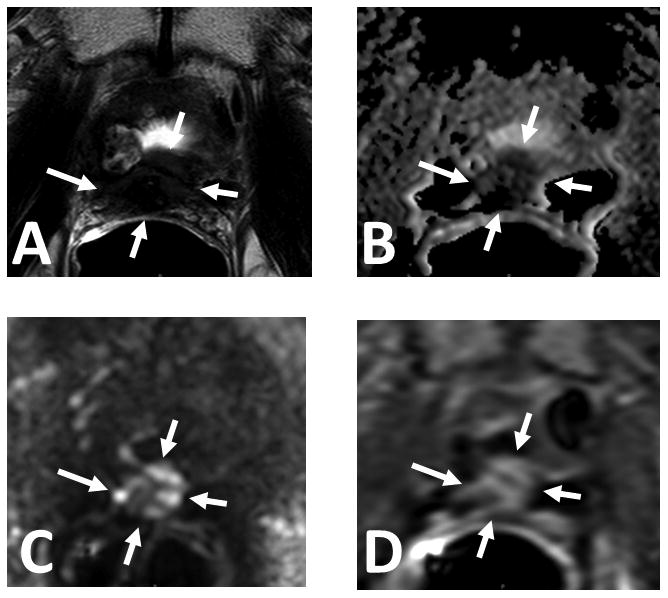
Figure 1.

61 year old male with serum PSA = 16.39ng/mL and S/P radical prostatectomy. Axial T2W MRI (A), ADC map of DW MR (B), b2000 DW MRI (C) and DCE MRI (D) shows a soft tissue lesion in the prostatectomy bed within the anastomosis (arrows). Targeted biopsy revealed recurrent prostate cancer.
