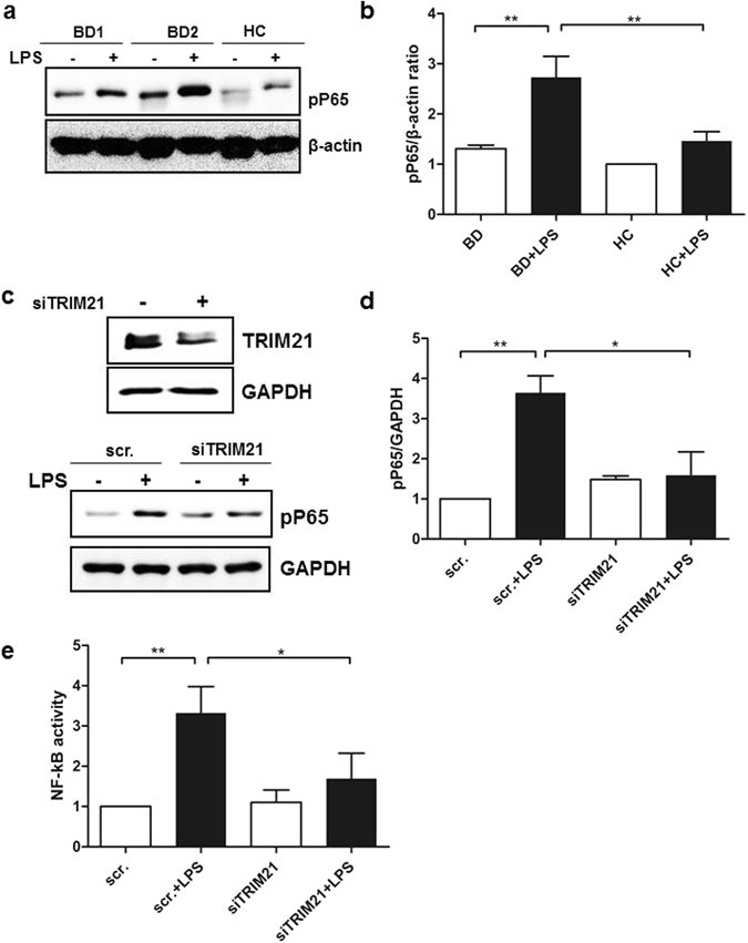
Figure 4.

LPS-stimulated BD monocytes exhibited enhanced nuclear translocation of NF-kB. (a and b) Monocytes isolated from healthy controls and BD patients were stimulated with LPS (1 µg/ml) for 1 h. Cell lysates were evaluated by western blot for levels of phosphorylated p65. β-actin was used as the loading control. A representative result from four independent experiments is shown in (a). (c and d) THP-1 cells were transfected with scrambled RNA or TRIM21 siRNA (10 nM) for 24 h, and the transfected cells were stimulated with LPS (1 µg/ml) for 1 h. (e) THP-1 cells were transfected with scrambled RNA or TRIM21 siRNA (10 nM) for 24 h and were incubated with pNF-kB-Luc vector (500 ng) and pβ-galactosidase (500 ng) for 24 h. The transfected cells were stimulated with LPS (1 µg/ml) for 6 h. The results represent NF-kB luciferase activity, normalized for β-galactosidase. Data are represented as the mean ± SD. The cropped blots are displayed and full-length blots are shown in the Supplementary Information.
