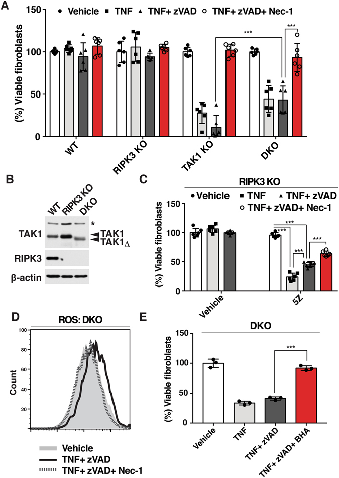
Figure 4.

TNF induces RIPK1-dependent noncanonical cell death independent of caspase activity and necroptosis in the absence of TAK1 (A) WT, Tak1-deficient (TAK1 KO), Ripk3 −/− (RIPK3 KO) and Tak1-deficient Ripk3−/− (DKO) were pre-treated with vehicle (DMSO) or 20 μM zVAD +/− 50 µM Nec-1 for 1 h and stimulated with 20 ng/ml TNF for 24 h. Cell viability was determined by crystal violet assay. Data are the result of 3 independent experiments and show mean percentages +/− SD. NS, not significant; ***p-value < 0.001; two-way ANOVA. (B) WT, RIPK3 KO and DKO fibroblasts were analyzed by immunoblotting. Tak1-deficient fibroblasts expressed a truncated nonfunctional form of TAK1 (TAK1Δ). β-actin is shown as a loading control. (C) RIPK3 KO fibroblasts were pretreated with 20 μM zVAD, 50 µM Nec-1 and/or 5Z-7-oxozeaenol for 1 h and stimulated with 20 ng/ml TNF for 24 h. Data are the result of at least 3 independent experiments and show mean percentages +/− SD. NS, not significant; ***p-value < 0.001; one-way ANOVA. (D) DKO fibroblast were pretreated with 20 µM zVAD and/or 50 µM Nec-1 for 1 h and stimulated with 20 ng/ml TNF. ROS were analyzed by CM-H2DCFDA at 6 h post TNF stimulation. The result shown is representative result of 3 independent experiments. (E) DKO fibroblasts were pre treated with 100 µM BHA and/or 20 µM zVAD for 1 h and stimulated with 20 ng/ml TNF for 24 h. Cell viability was determined by crystal violet assay. Data are the result of 3 independent experiments and show mean percentages +/− SD. ***p-value < 0.001; one-way ANOVA.
