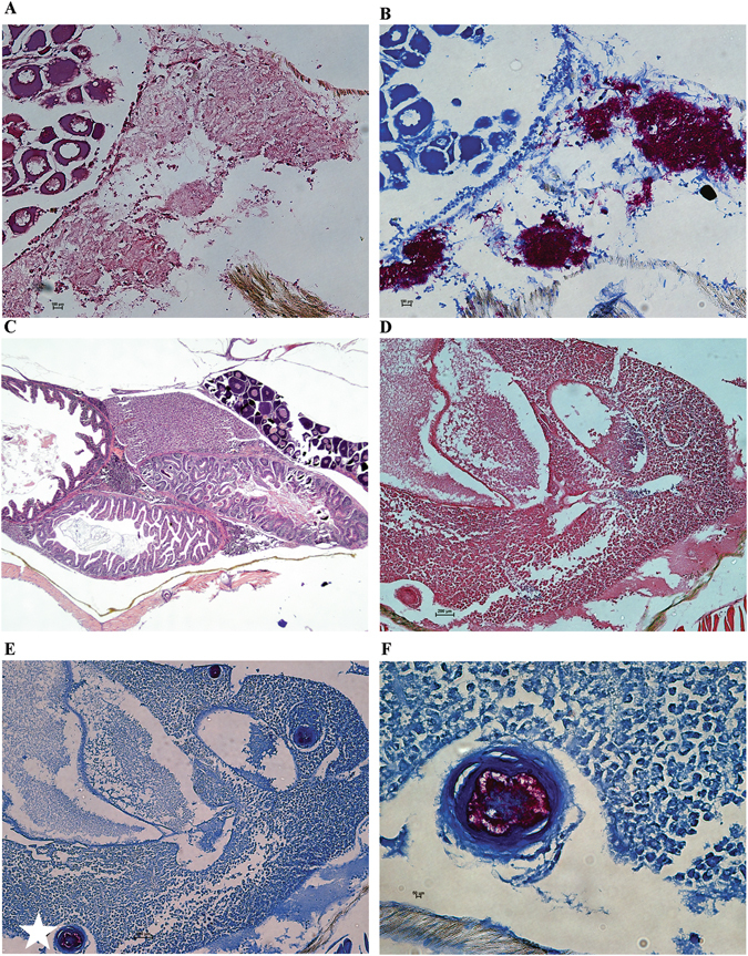
Figure 4.

M. marinum ΔwhiB4 caused less severe pathology. Histological analysis of fish tissues from the experiment described in Fig. 3A. (A,B) Liver sections of fish infected with WT M. marinum at 3 weeks post-challenge. Samples were analyzed with hematoxylin and eosin (H&E) staining (A) and Ziehl-Neelsen method of acid fast staining (B). Both are 20× magnification. (C) H&E staining of liver sections of fish infected with ΔwhiB4 at 3 weeks post-infection (4× magnification). (D,E) Liver sections of fish infected with ΔwhiB4 at 3 weeks post-infection (10× magnification) and stained with H&E (D) or Ziehl-Neelsen (E). The well-organized granuloma observed in (E, white star) was zoomed in for better visualization (F), 40× magnification.
