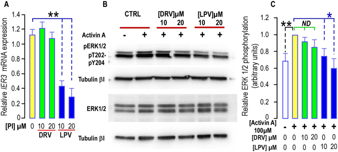
Figure 4.

Long term treatment with PIs impairs IER3 expression and ERK1/2 phosphorylation in hMADS cells. (A) Effect of PIs on IER3 expression. IER3 expression was determined in hMADS cells submitted or not to increasing doses of DRV (green bars) or LPV (blue bars) for 35 days. The analysis findings were assessed by real-time RT-PCR and normalized for the expression of 36B4 mRNA. Mean ± SEM was representative of three independent experiments. (*p < 0.05 and **p < 0.01). (B) PIs alter Activin A -induced ERK1/2 phosphorylation. Thirty µg of proteins prepared from hMADS cells treated with DRV or LPV for 35 days were loaded onto a 10% acrylamide gel. Sub-confluent cells were stimulated or not with 100 ng/ml activin A after 3 days of serum deprivation. Total or phosphorylated forms of ERK1/2 were assessed with specific antibodies. Activin A induced an increased phosphorylation of ERK1/2 that was blunted in cells treated with LPV. Expression of Tubulin-βI used as a loading control (lower panel). A representative Western blot is shown. (C) Quantification of the signals. The activin A-induced phosphorylation of ERK1/2 was measured using the Quantity one Program and compared to the expression of Tubulin-βI. Four and six determinations were analyzed when cells were treated respectively with DRV and LPV. Analysis was performed taking the unstimulated control as reference (black stars) or the activin A stimulated condition (green and blue stars) (*p < 0.05 and **p < 0.01). ND means “no significant difference”.
