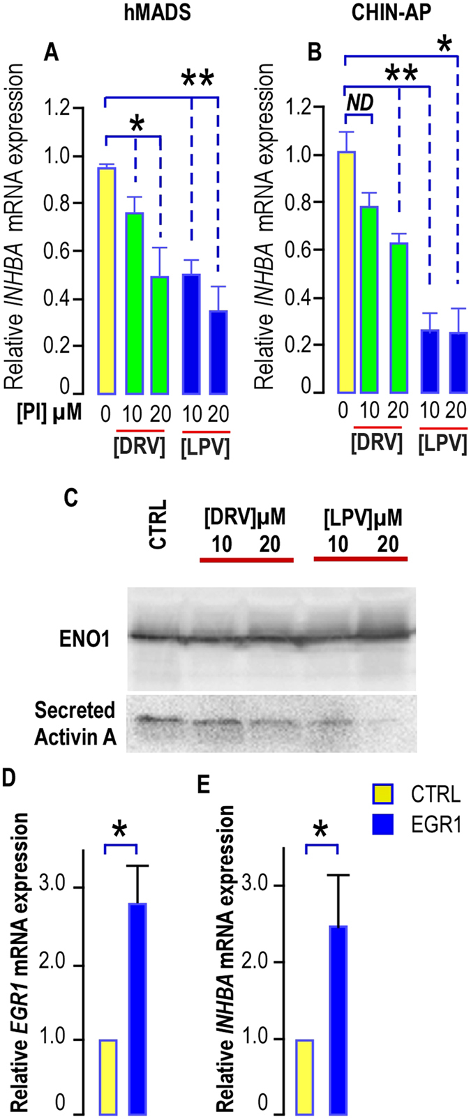
Figure 6.

LPV impairs INHBA expression and activin A secretion. (A,B) Expression of INHBA in hMADS cells and in chin-AP treated with PIs. INHBA expression was determined in hMADS cells and chin APs submitted or not to increasing doses of DRV (green bars) or LPV (blue bars) for 5 days. The analysis findings were assessed by real-time RT-PCR and normalized for the expression of 36B4 mRNA. Mean ± SEM was representative of four (hMADS) or three (chin-AP) independent experiments (ND = not significantly different, *p < 0.05, **p < 0.01, ***p < 0.001). (C) Effects of HIV-PIs on activin A secretion. Culture media were collected after a 4-day treatment of hMADS cells with or without PIs, filtered on 0.45 µM membranes, and concentrated with Amicon ultra-15 columns (NMWL, three KDa; Millipore). Activin A secretion was analyzed by Western blot performed under non reducing conditions because anti–activin A antibody selectively binds to the dimeric form of activin A. A representative Western blot showing the expression of Enolase 1 (ENO1) used as a loading control (upper panel) and of activin A (Lower panel) is presented. (D,E) Effect of EGR1 overexpression on INHBA. EGR1 and INHBA expressions were determined in hMADS cells transduced with a lentivirus allowing EGR1 expression. The analysis findings were assessed by real-time RT-PCR and normalized for the expression of 36B4 mRNA. Mean ± SEM was representative of four independent experiments. (*p < 0.05)
