
Figure 3.
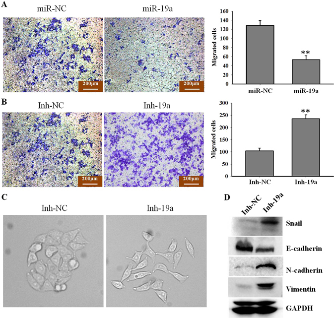
miR-19a contributes to EMT in NSCLC cells (A). A549 cells were treated with gefitinib (5 μM) and then transfected with miR-19a or miR-NC; after 24 h, cell migration was detected with a transwell assay. Migrated A549 cells were stained with crystal violet (left); miR-19a overexpression significantly decreased cell migration; quantified data are shown on the right. **p < 0.01; (B) A549 cells were treated with gefitinib (5 μM) and then transfected with Inh-19a or Inh-NC; after 24 h, cell migration was detected with a transwell assay, and migrated A549 cells were stained with crystal violet (left); miR-19a down-regulation significantly increased cell migration; quantified data are shown on the right. **p < 0.01; (C) A549 cells were treated with gefitinib (5 μM) and then transfected with Inh-19a or Inh-NC; the cell morphology is shown. The Inh-19a group exhibited spindle-shaped cells and loss of cell-cell contact, which suggested EMT; (D) A549 cells were treated with gefitinib (5 μM) and then transfected with Inh-19a or Inh-NC, and EMT pathway markers were detected by western blotting.