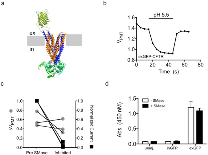
Figure 3.

Channel internalization does not underlie SMase-mediated inhibition of CFTR: (a) Molecular model of the exGFP-CFTR protein showing the GFP tag on the extracellular side of the plasma membrane (represented in grey). (b) Fluorescence measured from oocytes expressing exGFP-CFTR was sensitive to a drop in extracellular pH. Representative background-subtracted voltage from the photomultiplier tube during a brief exposure to pH 5.5 recording solution is shown. (c) Treatment with 10 μg/mL SMase for 10 minutes led to inhibition of currents (p = 0.0012) while the pH-dependent fluorescence change was not significantly affected (p = 0.4437, paired t-test). (d) In a cell-ELISA experiment, treatment with 10 μg/mL SMase for 10 minutes did not change the accessibility of the extracellular GFP on exGFP-CFTR to an anti-GFP antibody. Uninjected cells and cells expressing inGFP-CFTR showed minimal signal (n = 3).
