Figure 3.

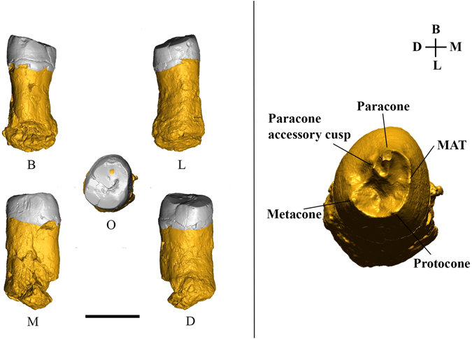
3D digital model of specimen EQH2, an upper right third molar. Left: Various views—B, buccal; L, lingual; M, mesial; D, distal; O, occlusal. The black bar represents 1 cm. Right: The enamel-dentine junction (EDJ) surface of EQH2.

3D digital model of specimen EQH2, an upper right third molar. Left: Various views—B, buccal; L, lingual; M, mesial; D, distal; O, occlusal. The black bar represents 1 cm. Right: The enamel-dentine junction (EDJ) surface of EQH2.