
Figure 2.
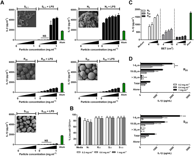
Hydroxyapatite particle size and shape dictates cytokine production by murine BMDCs. BMDCs (0.625 × 10⁶ cells · ml−1) from C57BL/6 mice were stimulated with concentrations ranging from 0.1 mg · ml−1 to 1 mg · ml−1 alone or after priming with LPS (1 ng · ml−1) for 3 h. Supernatants were collected 24 hours later and tested for IL-1β (A,C,D) by ELISA. Unprimed cells were collected and stained with LIVE/DEAD® fixable Aqua dead cell stain and CD11c+ and cell death was assessed by flow cytometry (B). Representative SEM pictures of HA particles (A). Results are mean cytokine concentrations (±SD) for triplicate samples ((A) vs LPS alone; (D) vs Total, NS p > 0.05, *p < 0.05 and ***p < 0.001). Data are representative of three independent experiments. (B) Results are pooled from three independent experiments. Error bars show means ± SEM for triplicate samples (vs media *p < 0.05); NS: non-significant.