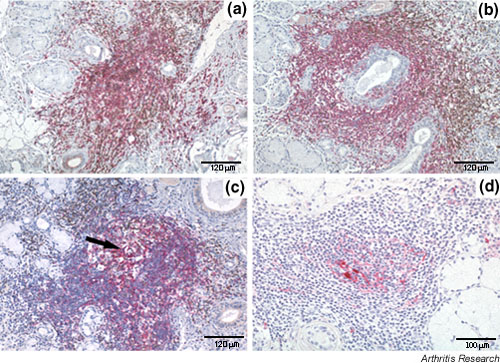
Figure 4.

Degree of lymphoid organization of the periductal lymphocytic infiltrates in salivary gland of patients with Sjögren's syndrome (SS). Paraffin-embedded sections were double-stained for CD3 (brown) and CD20 (purple) (a–c) and single-stained with CD21 (d). Inflammatory foci were classified as nonsegregated when T and B lymphocytes were not compartmentalized in distinct areas (a), as segregated in the presence of evident compartmentalization of T and B cells (b), and as segregated with germinal-centre-like structures (arrow) when a clear histological appearance (c) and networks of CD21+ follicular dendritic cells (d) were observed. Original magnification × 200.
