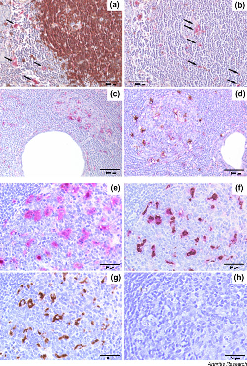
Figure 5.

Relationship between IL-18 expression and B-/T-cell compartmentalization (a,b) and germinal-center-like (GC-like) structures (c,d) in the salivary glands of patient with Sjögren's syndrome (SS) and, for comparison, in normal lymph nodes (e–h). Representative section of a large segregated aggregate double-stained for CD20 (brown) and IL-18 (purple) (a), and (b) sequential section with an irrelevant antibody replacing the anti-CD20, demonstrating the presence of IL-18-producing cells both in the T-cell (a, arrows) and B-cell (b, arrows) areas. (c) Single staining for IL-18, demonstrating a large number of IL-18-producing cells within ectopic GC-like structures in salivary gland from SS. (d) Double immunohistochemical staining for CD68 (brown) and IL-18 (purple), demonstrating the exclusive co-localization of IL-18 with CD68 macrophages. (e–h) An identical pattern of distribution in terms of IL-18 expression and co-localization with CD68 macrophages was observed in GCs of secondary lymphoid organs. Histomorphological analysis of the IL-18 positive cells within the GC showed evidence of engulfed apoptotic bodies in the cytoplasm (e) that identifies these cells as tingible body macrophages (TBMs). (f) Double immunohistochemical staining for CD68/IL-18 confirmed the exclusive co-localization of IL-18 with TBMs within the GC. Sequential sections in which the anti-CD68 (e), anti-IL-18 (g), or both the primary antibodies (h) were replaced with an isotype-matched irrelevant antibody confirmed the specificity of the double staining (h, negative control). Original magnification (a–d) × 200, (e-h) × 400
