
Figure 3. Role of MKP-1 in Monocyte Adhesions, Chemotaxis and The Molecular Mechanisms of Monocyte Priming by Metabolic Stress.
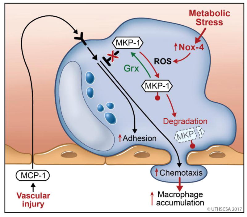
Chemoattractants, such as MCP-1 released by the vessel wall activate of monocytes (blue) in the blood stream and initiate monocyte recruitment in response to vascular injury. Monocyte transmigration and involves adhesion to and chemotaxis through the endothelial layer (pink). Both processes are initiated by protein kinase-dependent signaling pathways and counter-regulated by protein phosphatases (MKP-1). MKP-1 is sensitive to S-glutathionylation (
 ) of the catalytic cysteine residue which is reversed by the thiol transferase glutaredoxin (Grx). Metabolic disorders induce the expression of Nox4 in monocytes, an enzyme that generates reactive oxygen species (ROS) capable of catalyzing the S-glutathionylation of MKP-1. In the absence of adequate antioxidant protection, S-glutathionylated MKP-1 is rapidly degraded, promoting in MKP-1 deficiency, hyper-activation of monocyte adhesion and migration and subsequently the increased accumulation of monocyte-derived macrophages in the injured vessel wall.