
Figure 5. Genome therapy targeting at MAN2A1-FER breakpoint.
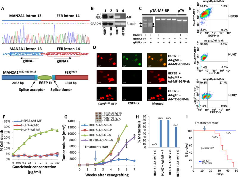
(A) Design of gRNA and recombination donor adenoviruses for MAN2A1-FER fusion gene. Upper panel: Sanger sequencing diagram of MAN2A1-FER chromosome breakpoint of HUH7 cells; Middle panel: Design of gRNA for pAD5-Cas9D10A-gRNAMAN2A1int13-gRNAFERint14; Lower panel: Design of homologous DNA sequences and EGFP-tk for pAD-MAN2A1int13-EGFP-tk-FERint14. The splicing acceptor and donor sequences correspond to the juncture sequences of intron13-exon 14 of MAN2A1and exon15-intron 15 of FER. (B) Expression of MAN2A1-FER in HUH7 cells. Lanes 1 and 2: immunoblots of protein extracts from HUH7 and HEP3B cells with antibodies specific for FER or GAPDH. MAN2A1-FER (MF) and FER protein are indicated. Lanes 3 and 4: RT-PCR of RNA from HUH7 and HEP3B cells with primers specific for MAN2A1-FER (MF) or β-actin. (C) In vitro cleavage assays were performed on BamH1 linearized pTA-MAN2A1int13-FERint14 vector using recombinant Cas9, S. pyogenes and in vitro transcribed gRNA− or gRNA+ as indicated. The cleavage generated 2446 and 1944 bp fragments of pTA-MAN2A1int13-FERint14 vector for gRNA−, and 2484 and 1906 bp for gRNA+. (D) Infection of HUH7 or HEP3B cells led to expression of EGFP-tk in HUH7 but not HEP3B cells. HUH7 and HEP3B cells were infected with pAD5-Cas9D10A-gRNAMAN2A1int13-gRNAFERint14 (ad-gMF) and pAD-MAN2A1int13-EGFP-tk-FERint14 (Ad-MF-EGFP-tk). Expression of Cas9D10A-RFP is indicated by red fluorescence, while expression EGFP-tk is indicated by green. HUH7 cells infected with pAD5-Cas9D10A-gRNATMEM135int13-gRNACCDC67int9 (Ad-gTC) and pAD-TMEM135int13-EGFP-tk-CCDC67int9 (Ad-TC-EGFP-tk) were used as specificity control. Selected images were shown. (E) Quantification of EGFP-tk integration/expression by flow cytometry as of (D). (F) Killing of HUH7 cells with ganciclovir. HUH7 or HEP3B cells were infected with pAD5-Cas9D10A-gRNAMAN2A1int13-gRNAFERint14/pAD-MAN2A1int13-EGFP-tk-FERint14 (Ad-MF). These cells were then incubated with various concentrations of ganciclovir for 24 hours. Cell deaths were then quantified with phycoerythrin labeled Annexin V through flow cytometer. HUH7 cells infected with pAD5-Cas9D10A-gRNATMEM135int13-gRNACCDC67int9/pAD-TMEM135int13-EGFP-tk-CCDC67int9 (Ad-TC) were used as specificity controls. (G) HUH7 and HEP3B cells were xenografted into the subcutaneous regions of SCID mice. These tumors were allowed to grow for 2 weeks before the treatment. These mice were treated with the indicated viruses plus ganciclovir (G, 80mg/kg) or PBS (P). The indicated drugs were applied through intraperitoneal injections 3 times a week until all the mice from control treatments died off. The tumor volumes were measured weekly. (H) Mice treated with MAN2A1-FER breakpoint therapy are free of cancer metastasis. (I) Mice treated MAN2A1-FER breakpoint therapy had no mortality.