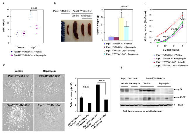
Figure 5. Rapamycin treatment mitigates MPN in Ptpn11E76K/+ mice.
(A) WBC counts in the peripheral blood of Ptpn11E76K/+/Mx1-Cre+ mice were determined before and after 12–14 weeks of treatment with Rapamycin (10 mg/kg body weight, once daily) or the vehicle (n=5/group). (B) Spleen weights were determined after 12–14 weeks of treatment with Rapamycin or vehicle. (C) BM cells were harvested from Ptpn11+/+/Mx1-Cre+ and Ptpn11E76K/+/Mx1-Cre+ mice after 12–14 weeks of treatment with Rapamycin or vehicle. These cells were assessed by CFU assays at the indicated GM-CSF concentrations. Experiments were performed three times with similar results obtained in each. Data shown are mean±S.D. from one experiment in triplicates. (D) Representative colonies growing in the medium containing GM-CSF (0.1 ng/mL) are shown. The average number of the cells per colony was determined by dividing the total number of the cells harvested from each plate with the number of the colonies in that plate (n=3/group). Experiments were performed in three pairs of mice with similar results obtained in each. Data shown are mean±S.D. from one experiment in triplicates. (E) Spleen lysates were prepared from Ptpn11+/+/Mx1-Cre+ and Ptpn11E76K/+/Mx1-Cre+ mice after 12–14 weeks of treatment with Rapamycin or vehicle. Levels of p-S6, p-4E-BP1, and Shp2 were determined by immunoblotting analyses. Each lane represents an individual mouse.

