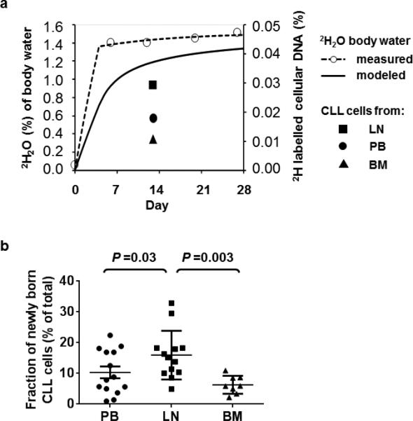
Figure 1. 2H2O enrichment in body water and 2H incorporation into cellular DNA to label “newly born” CLL cells.

(a) Heavy water enrichment in body water (left y-axis) and the fraction of 2H labelled cellular DNA (right y-axis) in CLL cells obtained from three anatomic compartments (LN, lymph node, PB, peripheral blood, BM, bone marrow) in a representative patient (HW04) is shown. 2H2O was consumed for 28 days. On days 7, 14, 21, and 28 saliva and/or serum samples were collected to determine 2H exposure. These time points (open circles, dashed line) were used to calculate the average 2H exposure per day (solid line). The goal range of 1 to 1.5% 2H2O in body water was achieved by day 7. (b) The fraction of newly born CLL cells on day 13 is shown for PB (circle), LN (square), and BM (triangle). Plot depicts the mean ± standard error of the mean. Comparisons between different anatomic sites were done by repeated measures ANOVA.
