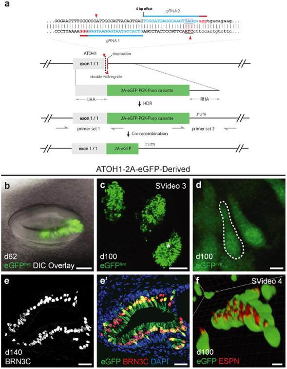
Figure 3.

Development of an ATOH1 fluorescent reporter hESC line for tracking hair cell induction. a, ATOH1-2A-eGFP CRISPR design. The two guide RNAs (blue, with PAM sequence in red) direct Cas9n to make two nicks (red triangles) near the stop codon (underlined with pink background) of ATOH1. The resulting DNA double strand break is repaired by the donor vector, which has a 2A-eGFP-PGK-Puro cassette and 1kb left and right homology arms (LHA and RHA). The LoxP-flanked PGK-Puro sub-cassette is subsequently removed by Cre recombinase. In ATOH1 expressing hair cells, eGFP is transcribed along with ATOH1. b-d, Representative live cell images of eGFP+ hair cells in 62- and 100-day-old inner ear organoids. The asterisk in panel (c) denotes the approximate location of the hair cells in panel (d). e, Expression of BRN3C in 140-day-old eGFP+ hair cells. f, Expression of ESPN in the hair bundles of 100-day-old eGFP+ hair cells. Scale bars, 100 μm (c), 50 μm (b), 25 μm (e), 5 μm (d, f).
