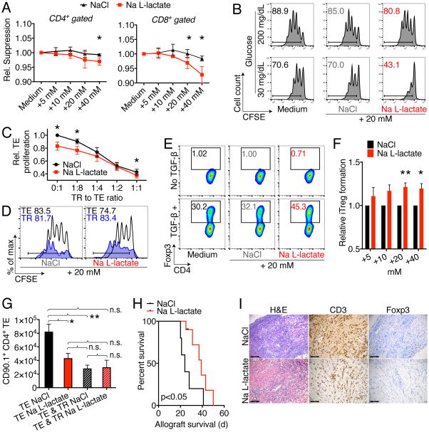
Figure 5. L-lactate impairs T effector cells but not Tregs.
(A) C57BL/6 splenocytes were labeled with CFSE and stimulated with 1 μg × ml−1 soluble CD3ε mAb, and CD4+ as well as CD8+ T cell proliferation was assessed after three days. Incremental doses of Na L-lactate suppressed T cell proliferation compared to NaCl control. Data pooled from four independent experiments (paired Student t-test). (B) CD4+CD25− Tconv were CFSE labeled and stimulated with anti-CD3ε mAb and irradiated antigen presenting cells. Reduced ambient glucose augmented the suppressive effect of pH neutral Na L-lactate suppression of T cell proliferation. Data representative of three independent experiments. (C) CFSE labeled effector T cells (TE) were co-stimulated with anti-CD3ε mAb and irradiated CD90.2− antigen presenting cells, and suppressed by adding regulatory T cells (TR) at the indicated ratios. In contrast to TE proliferation, TR suppressive function is not impaired by added Na L-lactate (7/group, paired Student t-test). (D) Treg suppression assay with CD90.1 Treg and CD90.2 effector T cells to track proliferation of each subpopulation. Na L-lactate impaired Teff proliferation, but not TR. Data representative of three independent experiments. (E) Representative and (F) quantified data from 10 independent Treg induction experiments under the same conditions as in Fig 1A (paired Student t-test). Added pH neutral Na L-lactate augments induced Treg formation. (G) B6/Rag1−/− mice were adoptively transferred with 1 × 10⁶ Tconv ± 2.5 × 10⁵ Treg cells, and treated i.p. with 60 μl × g−1 of 150 mM Na L-lactate or NaCl control for seven days. Administration of Na L-lactate reduced TE (CD90.1+CD4+) homeostatic proliferation without affecting TR suppressive function. * and ** indicates p<0.05 and p<0.01, respectively (3–11/group, ANOVA with Tukey’s multiple comparisons test). (H, I): Cardiac allografts from BALB/c (H-2d) donors were transplanted into the abdomens of MHC-mismatched C57BL/6 (H-2b) recipients. Recipients were then treated for 14 days with 0.2 mg × kg−1 × d−1 rapamycin i.p., and 60 μl × g−1 body weight of 150 mM Na L-lactate (9 μmol × g−1 × d−1, 10/group) or NaCl (5/group) control. (H) Na L-lactate injections led to a brief prolongation of allograft survival (Mantel Cox test). (I) Allograft histology at 20–28 d post-transplant shows a similar degree of lymphocyte infiltration, with equal CD3 and Foxp3 cells (4–5/group). Error bars indicate SEM.

