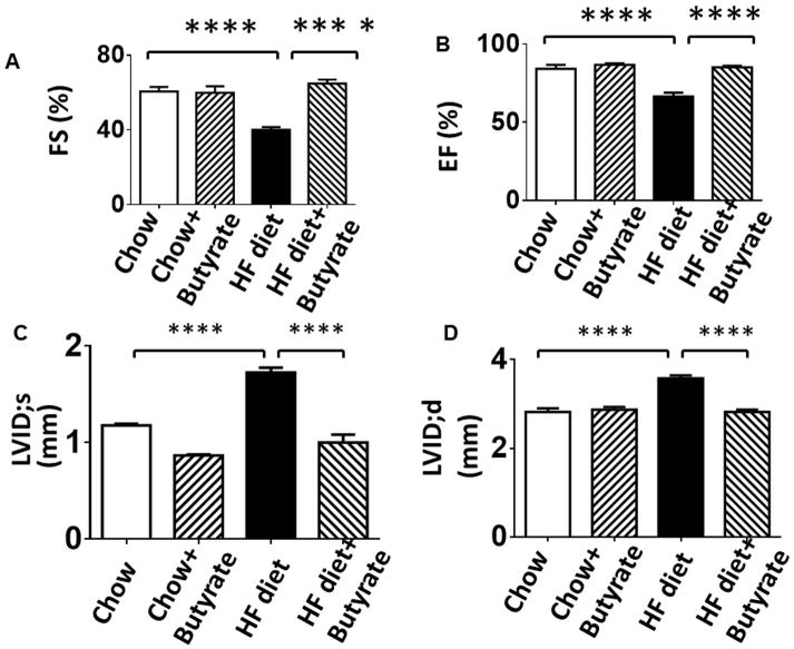
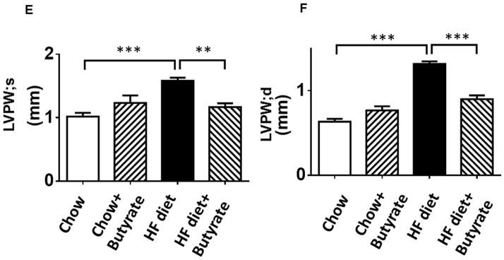
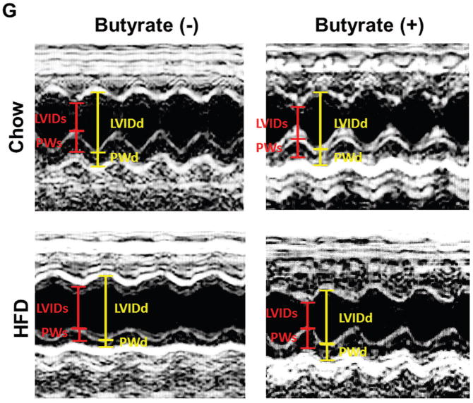
Figure 3. Sodium butyrate treatment improved impaired left ventricular function in HFD-fed mice.
Echocardiographic measurements of ventricular functional parameters includes A: Fractional Shortening (FS) B: Ejection Fraction (EF); C. Left Ventricular Internal Dimension in Diastole (LVID;d); D: Left Ventricular Internal Dimension in Systole (LVID;s); E and F: Left ventricular posterior wall (LVPW;s and LVPW;d); G: Representative image of M-mode. Data are shown as means ± SEM (n=4–5 per group). ** p<0.01, ***p<0.001, ****p<0.0001.


